Anomaly Scan

By the 20th week of pregnancy, the realization completely sinks in that you are going to be a mother! Congratulations for carrying your baby in your tummy safely for so long and creating a nurturing environment for your little one. But your responsibility does not end here. In fact, it is just getting started.
Dr. Vishwas Khichar who have special expertise and exhaustive in-depth experience of Sonologist and Radiologist.
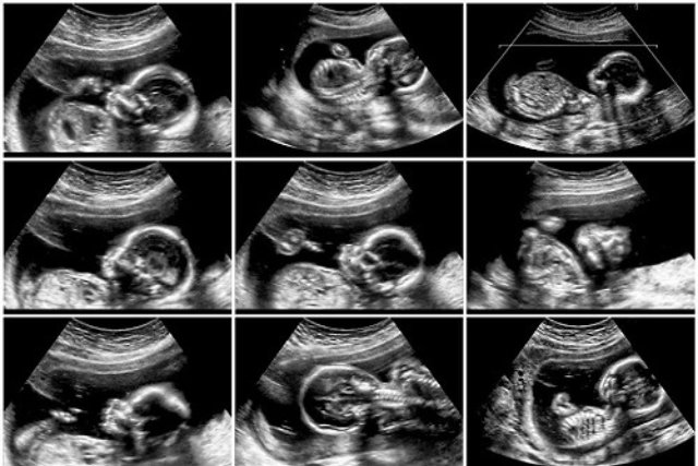
The scan aims to look for any major physical abnormalities in the growing baby. The scan can be as a dating scan where a black and white 2-dimensional (2-D) image is produced which gives the side-view of the baby in the womb. This image shows the baby’s face and hands at 20 weeks and gives the healthcare specialist (a sonographer) an idea of what is going inside. This can be undoubtedly exciting!

What is Anomaly Scan?
Anomaly Scan or mid-pregnancy scan is an ultrasound scan done between the 18th and 21st week of pregnancy to take a closer look at the baby and the womb (uterus) and to have an idea where the placenta is lying.
Why is Anomaly Scan done?
The mid-pregnancy anomaly scan is done for checking any physical abnormalities in the growing baby. Although it can’t pick up every problem, it gives the healthcare specialist (a sonographer) an idea about the baby’s bones, heart, brain, spinal cord, face, kidneys and abdomen and allows the healthcare specialist identify the following conditions (some of which are very rare):
- Anencephaly
- Diaphragmatic hernia
- Gastroschisis
- Open spina bifida
- Bilateral renal agenesis
- Lethal skeletal dysplasia
- Edwards’ syndrome or T18
- Patau’s syndrome or T13
- Cleft lip
- Serious cardiac abnormalities
Mostly the scan shows that the baby is developing normally, but in a few cases, the sonographer will find or suspect a problem.
How is it done?
A sonographer asks the patient to lie on a couch and uncover the abdomen and applies gel on the abdomen. Then he/she passes a handheld probe over the skin of the abdomen to examine the baby’s body. The gel is applied to make sure that there is good contact between the probe and the skin. As the probe moves, a black and white 2-D image of the baby will appear on the ultrasound screen. For a better view, the sonographer will ask the patient to drink water to have a full bladder before the appointment. At times the sonographer may apply slightly more pressure to get a better view of the baby.
The entire process of mid-pregnancy scan takes only around 30 minutes.
